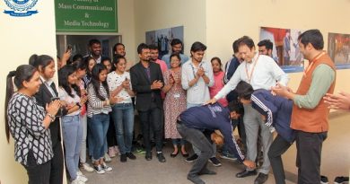
The Savant Association, Department of Physics, FOSC, organizes an industrial visit
SGT Times Reporter
Gurugram, May 2
The Savant Association, Department of Physics, FOSC, organized an industrial visit to the Sophisticated Analytical Instrument Facility (SAIF), AIIMS, New Delhi.
The students from the Department of Chemistry, Forensic Science, Environmental Science, and Physics were taken to the facility.
The Department of Science and Technology (DST), Government of India, established a Regional Electron Microscope facility at AIIMS under its RSIC/SIF program. This center is presently known as Sophisticated Analytical Instrument Facility (SAIF) – AIIMS, New Delhi, under the SAIF Program of DST.
DST has set up Sophisticated Analytical Instrument Facilities SAIFs in different parts of the country under its Sophisticated Analytical Instrument Facilities Programme to provide the facilities of sophisticated analytical instruments to the research workers in general and especially from the institutions which do not have access to such instruments to enable them to pursue R&D activities requiring such facilities and keep pace with developments taking place globally. The instrument facilities provided by the SAIFs are being utilized by 8,000 users every year from academic institutions, R&D laboratories, and industries from all over the country.
OBJECTIVES OF THE SAIF:
To carry out analysis of samples received from the scientists/institutes; To provide facilities of sophisticated analytical instruments to scientists and other users from academic institutes, R&D laboratories, and industries to enable them to carry out measurements for R&D work; To acquire and develop the capability for preventive maintenance and repair of sophisticated instruments;
After reaching there, the technical staff introduced the students to biological sample preparation for TEM and Fixation. Fixation is a vital step in preparing biological specimens for electron microscopy. It is a process that prevents autolysis, changes in volume and shape, and preserves the chemical constituents of the cell.
The students were also briefed about the following facilities and visited different labs to see and learn about them.
ULTRAMICROTOME
Ultramicrotomy is a method for cutting specimens into extremely thin slices, called ultra-thin sections, that can be studied and documented at different magnifications in a transmission electron microscope (TEM). It is used mainly for biological specimens, but sections of plastics and soft metals can also be prepared.
GLASS KNIFE MAKER
Glass knives can be produced by hand using pliers with two raised bumps on one jaw and a single bump between the two bumps on the opposing jaw. Still, special machines called “knife-makers” are used in most electron microscopy laboratories to ensure repeatable results.
AUTOMATED TISSUE PROCESSOR
Tissue processing entails a series of processes that take human tissue from fixation to the point where it can be fully infiltrated with an appropriate histological wax and embedded, ready for microtome section cutting. Fixation, dehydration, cleaning, and embedding are the steps involved in tissue processing. Tissue-transfer machines and fluid-transfer machines are the two basic types of automated tissue processors. The tissue processor is used in histopathology laboratories to fix, dehydrate, clarify automatically, and infiltrate tissue samples with paraffin to prepare them for laboratory testing.
TALOS EDS FACILITY AT TEM
The TEM EDS analysis is an excellent technique for determining chemical composition. Elemental analysis is a technique that can be performed on both materials and metal samples. Although TEM EDS is not a quantitative approach, it may detect small amounts of elemental composition, which can be used to examine for contamination or faults.
HR-TEM (TECNO)
HRTEM (high-resolution transmission electron microscopy) is a TEM imaging mode that provides atomic-scale imaging of a sample’s crystallographic structure. It’s a useful instrument for studying nanoscale characteristics of crystalline materials because of its excellent resolution.
SEM
A scanning electron microscope (SEM) is a microscope that produces images of a sample by scanning the surface with a focused beam of electrons. The electrons interact with atoms in the sample, producing various signals that contain information about the surface topography and composition of the sample.
The students were accompanied by the Head of the Physics department, Dr. Mukesh Kumar, Dr. M.T. Beig, Advisor Savant Association, and Dr. Yogesh Sharma, Co-advisor Savant Association, Physics Department, FOSC.